


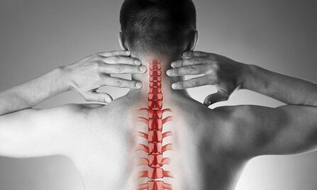
Cervical osteochondrosis or spondylosis occurs as a result of changes in the shape and structure of the vertebrae.Despite the fact that the cervical region is quite short compared to the total length of the spine, it is perhaps the most important part of the spine.Each pair of adjacent vertebrae forms intervertebral foramina through which nerve roots exit and are directed to every muscle and organ of the upper half of the body.The vital vessels providing blood supply to the brain pass through other openings - in the lateral processes of these vertebrae.
Causes of osteochondrosis of the cervical vertebrae
The causes of osteochondrosis are:
- injuries,
- "sedentary" work on a monitor located below eye level,
- physical labor associated with carrying heavy loads,
- driving a car for a long time,
- work "on the phone" without using remote devices (in this case, the operator presses the handset to his ear with his shoulder)
- constitutional features (torticollis, congenital changes in the cervical vertebrae, short neck)
Formation of pathological changes in the vertebrae
With osteochondrosis, small sharp points begin to form on the edges of the vertebral bodies, which can injure nearby structures.Most often, this happens in response to excessive loading of the cervical spine and is not only a result of the "aging" of the intervertebral joints (remember that previously osteochondrosis was considered a degenerative, natural "age" disease, like osteoarthritis).As the disease progresses, the vertebral endplates become denser and the height of the intervertebral discs decreases.These discs usually act as shock absorbers between the vertebrae and, among other things, prevent damage to the spinal roots.In progressive osteochondrosis, a protrusion (hernia) of the pulpous nucleus of the intervertebral disc occurs, on which, in the course of the disease, increasing pressure is exerted, while the ligaments that "hold" on all sides are weakened.This herniation can also compress spinal structures and cause neurological manifestations of the disease.
What are the symptoms of cervical osteochondrosis?
Osteochondrosis of the cervical vertebrae with pain syndrome
Any pain in the neck area makes one suspect pathology of the cervical spine.Depending on the increasing intensity of the pain syndrome, they are divided into 4 stages, in the first stage the patient feels numbness, tingling, a feeling of "tightness" in the area of a certain muscle group, in the fourth stage - the most severe - the pain is so intense that it leads to immobilization of the patient and loss of work capacity.
In addition to pain in the cervical and occipital regions, the patient notes "referred" (radiating) pain in the upper limb and subscapular lateral regions of the chest.
Osteochondrosis of the cervical vertebrae with radicular syndrome
The involvement of the nerve roots in the process is indicated when pain, numbness and tingling spread to the lower jaw, upper back, forearm and fingers.At the same time, the patient draws attention to the fact that he "seems to rest" on his arm and sleeps uncomfortably.There is morning stiffness in the joints of the fingers, which lasts no more than 10-15 minutes.With the development of radicular syndromes, a decrease in muscle strength of the upper limbs can be observed on examination.
Osteochondrosis of the cervical spine with vertebral artery syndrome.
The involvement of blood vessels in the process (compression by a hernial protrusion or osteophyte) is indicated when the patient complains of frequent attacks of headache, especially after being in a certain position for a long time, when throwing back the head (for example, when swimming breaststroke), if there is tinnitus and dizziness.This clinical situation is well identified using ultrasound (with "Doppler mapping mode").Ultrasound reveals distortion of the vertebral arteries and narrowing of their lumen.In this case, we can talk about surgery, because the pronounced change in blood flow in the spinal arteries is a risk factor for a stroke.
Osteochondrosis of the cervical vertebrae with "cardiac (cardiac) syndrome"
This syndrome forces the patient to consult a cardiologist first, since the main complaints refer to pain in the left half of the chest, the subscapular area, which weakens or intensifies when performing physical activity or changing the position of the body.After excluding myocardial infarction and other heart diseases, the patient is admitted under the supervision and treatment of a neurologist and orthopedist.
Diagnosis
To clarify the diagnosis, four methods are used: radiography, ultrasound, computed tomography and nuclear magnetic resonance.
The most accessible method is still radiography of the cervical vertebrae;X-ray in the lateral projection ("side view") is most informative.This method allows, with a first approximation, to determine the presence of injury and gross structural changes in the vertebrae.
An ultrasound examination (ultrasound) is performed to clarify the condition of the vertebral arteries.With the help of this method, it is determined whether the blood flow is disturbed and, if so, to what extent and what obstacles have occurred and where they are located.
Computed tomography (CT).It allows you to more accurately assess the state of bone structures, the degree of density of bone tissue and allows you to see smaller osteophytes (bone growths) than is possible with radiography.
Magnetic resonance imaging (MRI).This type of examination is indispensable if there is a suspicion of the presence of a hernia, the exact localization of the damage to the spinal cord and the degree of this damage.This research is necessary if the question of operative (surgical) treatment of diseases of the cervical spine is raised.
Treatment of cervical osteochondrosis
Drug treatment
The standard set of drugs for the treatment of cervical osteochondrosis reflects the goals of treatment: pain relief by eliminating painful muscle spasms and inflammation of the nerve roots, while increasing the mobility of the spine.To achieve these goals, they are mainly used by using pain relievers, NSAIDs - non-steroidal anti-inflammatory drugs, muscle relaxants.It should be remembered that self-medication with drugs from these groups can be dangerous, since there is a possibility of misinterpreting the symptoms, as well as underestimating the side effects of these drugs.Topical (skin) NSAIDs in the form of gels are widely used, and when the pain stops, these same drugs can be used in the form of ointments.
To treat osteochondrosis at a deeper, "basic" level, slow-acting systemic drugs are used.These substances restore the cartilaginous structures of the vertebrae and prevent their further damage.Treatment courses are long, the effect lasts for many months.
Cervical osteochondrosis has significant differences from the pathology of other parts of the spine.Pain in the neck area in this case can be provoked not by signals from the suffering spinal nerves, but by painful chronic muscle tension - all this together is called muscle-tonic syndrome.This is a completely "benign" condition that responds well to treatment with the same set of drugs: nonsteroidal anti-inflammatory drugs, muscle relaxants, use of intramuscular "blockades" using steroids.Usually, the doctor detects sharp pain when palpating the so-called "trigger" points along the entire cervical spine, as well as in the area of the muscles of the upper shoulder girdle.More often, this pathology occurs in women, most of whom are under the age of 40.Despite the severe pain syndrome, the neurovascular structures remain intact and the blood flow in the head area is not affected.
Manual therapy
This method of treatment can be effective for recent (often as a result of a minor injury, subluxation) neck pain that is not accompanied by dizziness or other changes in the nervous system and circulatory system.It is permissible to resort to manual therapy only after a thorough examination;in addition, the doctor who performs this procedure must have sufficient experience in the field of traumatology and orthopedics.For the "old" forms of the disease, the use of manual therapy is dangerous!
There are two known methods for this type of intervention:
- manipulation (sharp short blows with significant force, aimed at eliminating subluxations, the well-known "bone snaps");
- mobilization (the method is based on smooth stretching of the neck after warming up and relaxing the muscle corset of the neck).
A combined method based on a combination of two main ones is also used.It is important to remember that in addition to these contraindications, manual therapy is prohibited for any diseases accompanied by increased blood pressure, for any pathology of the thyroid gland and ENT organs.
Treatment of cervical osteochondrosis at home
Therapeutic exercises for cervical osteochondrosis
The first and foremost rule for beginners in physical therapy is not to perform exercises while overcoming painful sensations.It goes without saying that you shouldn't start in the "acute" period when the pain has just started.Another important recommendation is to avoid sudden movements and circular movements in the cervical spine.
Each session should begin with a short, light self-massage of the neck muscles.
This is followed by a "warm-up" warm-up:
- Arms are lowered along the body, shoulders are level, back is straight (you can check your posture by gently pressing your heels, shoulder blades and buttocks against the wall).We walk in place for 1 minute on the whole foot, 1 minute on the toes, 1 minute on the heels.
- The starting position is the same.We clench our hands into fists, raise and lower our shoulders, arms are straight.The movements are slow, we do 20 repetitions, the last lift is 5 seconds longer.We make sure that the neck muscles do not tighten.
- The starting position is the same.We tilt our heads one by one to the right, then to the left.The movements are smooth, one incline for 8 counts, at the end point of the incline - hold for 8 seconds.
- The starting position is the same or sitting on a hard chair.Smooth tilts of the head forward, at the end point - hold for 8 seconds
- The starting position is the same or sitting on a hard chair.Slowly tilt your head forward until your chin touches your chest, then slowly roll your head to the right (for 4 counts) and to the left (for 4 counts).Avoid muscle strain.
- The starting position is the same or sitting on a hard chair.We raise our shoulders for 4 counts, then smoothly lower them for 4 counts.10 repetitions.
- The starting position is the same or sitting on a hard chair.We raise our shoulders, but now we perform circular movements from front to back, for 8 counts.10 repetitions.
- We straighten the back and check our posture.For 4 counts, we collect the shoulder blades behind our back, trying to connect them, at the end point we hold for 8 seconds, after which we return to the starting position.
Pillows
As already mentioned, the hypertonicity of the neck muscles is the first and often the main reason for the development of cervical osteochondrosis.The rational selection of pillows and mattresses, ensuring a calm and comfortable position during sleep, are no less important than gymnastics, physiotherapy and medicines.
When choosing a mattress, pay attention to the composition of the filler (products that are at least half made of coconut husks, that is, with a sufficient degree of hardness, are suitable).Soft spring mattresses do not provide sufficient straightening of the spine.The most optimal sleeping position is on your side, with one or both knees pulled toward your stomach.The pillow should be positioned so that it fills the entire space between the shoulder, the ear and the mattress, while the parietal part (crown) of the head is in the same horizontal line with the spine.Pillows that are too high and too low, as well as soft, should be avoided.The ideal option is a product with an ergonomic shape, that is, in this case with a small pressure roller on one side.
General recommendations
Pay attention to your posture.When walking or standing, the correct position is when the chest is forward and the stomach is retracted.
Avoid sitting for long periods of time.A simple rule for the prevention of cervical osteochondrosis is known: after every 60 minutes of work, 10-15 minutes of walking or warming up is necessary.
The work chair should have a high headrest or backrest.
When sitting, your feet should rest on the floor and your neck should not be tense.For this purpose, use special orthopedic devices: supports under the neck when driving in a car, a pillow under the back.
Avoid heavy lifting.If necessary, drop to your knees, hold a heavy object on your torso, and then stand up smoothly, using the strength of your leg muscles, but not the "pull" of your back.
Do not bend over with straight legs.Use stands or work surfaces to bring the subject closer to you instead of leaning your face toward the subject.Try to do your homework while sitting on a chair or an exercise ball.
If you must use a mop, broom or rake, do not strain your arms, back, neck or bend sideways.
Avoid breaststroke.